Shortness of breath

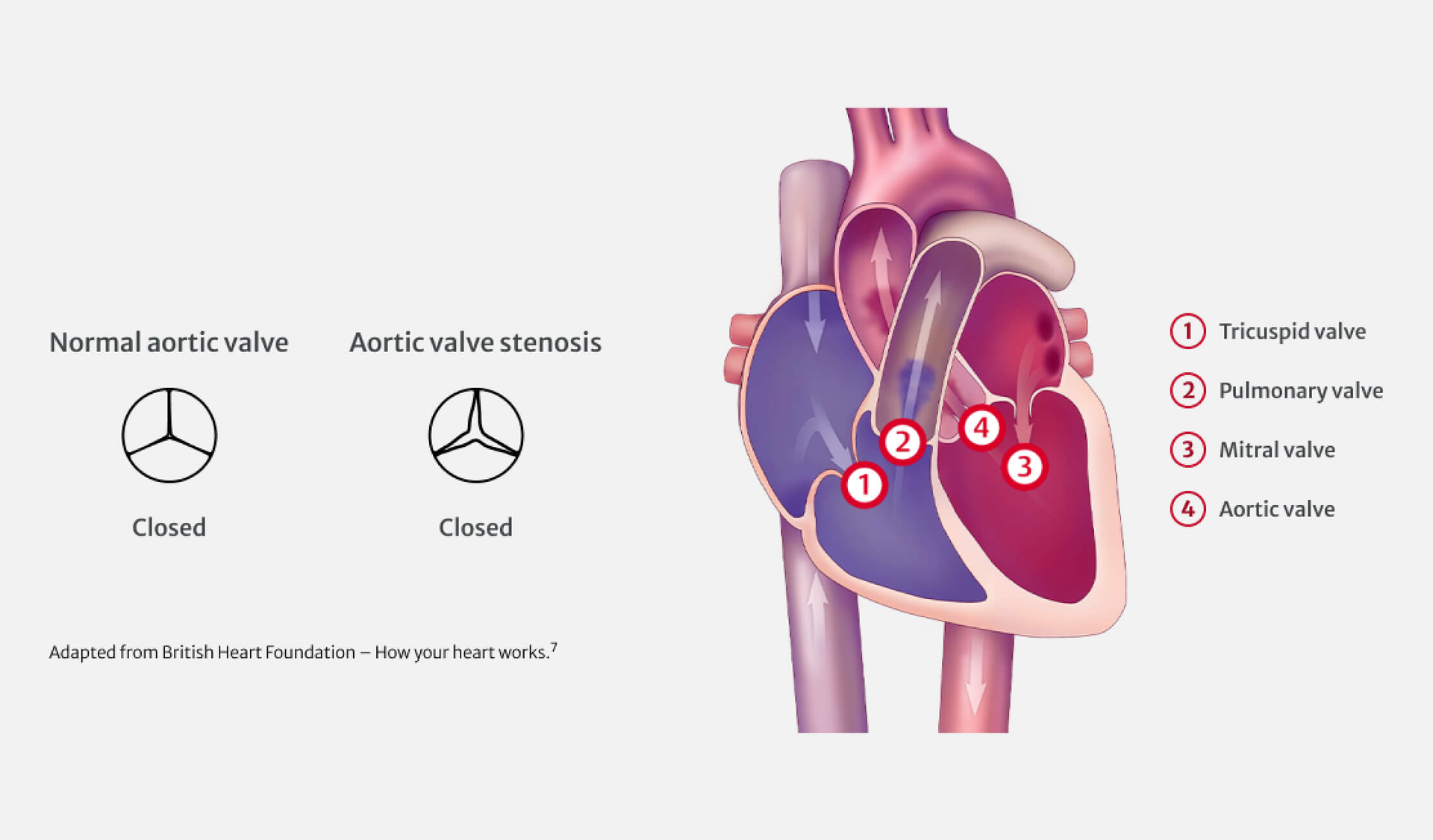
Valvular heart disease occurs when one or more valves in your heart is damaged or diseased and does not work properly.1 There are different types of valvular heart disease that can cause problems in any of your heart’s four valves. Aortic stenosis stands out as one of the most prevalent forms of valvular disease.2

You may not experience any symptoms at first, but as aortic stenosis progresses, your heart has to work harder to compensate for it. You may then experience:4,8

Shortness of breath

Chest pain
(also called angina)

Dizziness or
lightheadedness

Feeling faint
or fainting

Feeling tired, especially
after being active
Before you see a doctor or a cardiologist about your heart valve problem, you may have experienced some type of physical sign or discomfort.9
However, in some cases a heart valve problem may cause no symptoms at all,4 and your valve issues may be identified during a routine exam. Heart valve problems may produce a heart murmur, which can be heard through a stethoscope.9 If your doctor hears a heart murmur, particularly if it is new or loud, they will want to investigate further.
Cardiologists and surgeons have many ways of diagnosing heart valve disease.9 The most important is the echocardiogram.

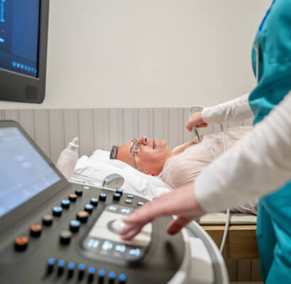
Echocardiography uses ultrasound to allow a cardiologist to see the function of your heart valves and the contractions of your heart muscle.9 During an echocardiogram, ultrasound waves are projected onto your heart and a computer translates the sound waves into an image.9 Echocardiography does not involve any radiation.
Most echocardiograms are performed by placing an ultrasound probe on your chest.10 This is called a “transthoracic” echocardiogram10 and only takes a few minutes.
In some cases, you may have a transesophageal echocardiogram.10 With this type of echocardiogram, the ultrasound probe is inserted into your oesophagus through your mouth.9 Because your oesophagus is very close to the back of your heart, it provides excellent images of the heart and its valves.
