Edwards Lifesciences Logo

Valve defects and diagnosis

placeholder
placeholder
placeholder

Causes of heart valve disease

Heart valves can fail by becoming narrowed (stenotic) so that they block the flow of blood or leaky (regurgitant) so that blood flows backward in the heart. Sometimes a valve is both stenotic and regurgitant. A variety of conditions can cause these heart valve abnormalities. Learn more about some of these conditions below.

Degenerative valve disease

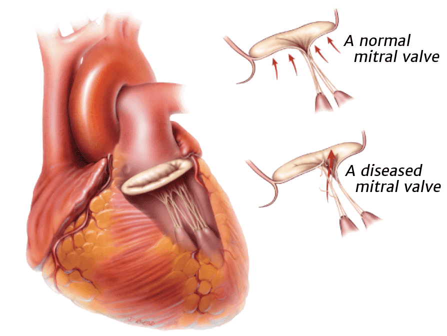
This is a common cause of valvular dysfunction. Most commonly affecting the mitral valve, it is a progressive process that represents slow degeneration from mitral valve prolapse (improper leaflet movement), a condition that affects 4-5 percent of the general population. Over time, the attachments of the valve thin out or rupture, and the leaflets become floppy and redundant. This leads to leakage through the valve.

Diagnosis of valve disease

Before a patient sees a primary care physician or a cardiologist (a doctor who specializes in understanding the heart) concerning a heart valve problem, he or she has often experienced some type of physical sign or discomfort. Some physical signs of heart valve disease include: angina (chest pain), tiredness, shortness of breath, lightheadedness or loss of consciousness.

However, in some cases a heart valve problem may cause no symptoms at all. These heart valve issues can often be identified by use of a stethoscope on routine physical examination. Heart valve abnormalities, whether stenosis or regurgitation, often produce a heart murmur. A heart murmur, particularly if it is new or loud, should prompt further investigation by your physician.

Cardiologists and surgeons have many ways of diagnosing heart valve disease. The most important is the echocardiogram.

echocardiography
echocardiography
echocardiography

Echocardiography

Echocardiography is a special application of ultrasound that enables the cardiologist to observe the function of your heart valves and the contractions of your heart muscle. During an echocardiogram, ultrasound waves are projected onto the heart. The reflected ultrasound is “captured” by a transducer, and a computer translates the sound waves into an image. Most echocardiograms are performed by placing the ultrasound probe on a person’s chest. This is called a “transthoracic” or “surface” echocardiogram. This test is non-invasive and takes only a few minutes.

In some cases, a tranesophageal echocardiogram is performed. With this type of echocardiogram, the ultrasound probe is inserted into the esophagus through the mouth. Because the esophagus is very close to the back of the heart, this sort of echocardiogram provides excellent images of the heart and its valves.

catherization
catherization
catherization

Catherization

Cardiac catheterization (angiography) helps to determine the function of the coronary arteries and the heart valves. Cardiac catheterization is the process by which a tube is inserted into the blood vessels and/or heart. The tube injects a contrast medium (dye) which is then visualized with X-rays. Coronary angiography is particularly useful for analyzing the coronary vessels of the heart. Most patients have a coronary angiogram, or cardiac catheterization, before heart valve surgery; this is necessary to determine whether any blocked coronary arteries require treatment at the time of surgery.

x-ray
x-ray
x-ray

Chest X-ray

A chest X-ray can be important in the detection of calcium deposits in the heart, such as on heart valves. It will also show the size and shape of the heart and lungs. All patients will have a chest X-ray before heart valve surgery.

couple consultation
couple consultation
couple consultation
consultation
consultation
consultation
There are options to treating diseased heart valves

Valve treatment

consultation
consultation
consultation
There are options to treating diseased heart valves

Diseased heart valves can be addressed in several ways.  If repair or replacement becomes necessary, there are treatment options to consider. A team of medical specialists will be committed to your safety and comfort before, during and after your procedure.

heart diagram
heart diagram
heart diagram
Maintaining a healthy heart is important to living a healthy life

What the heart does

heart diagram
heart diagram
heart diagram
Maintaining a healthy heart is important to living a healthy life

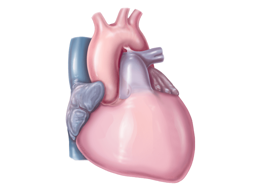
Beating about 2.5 billion times over an average lifetime, the heart provides blood flow to all the cells, tissues, and organs in your body when it's working properly. Clasp your two hands together and that's about the size of a healthy heart.  It's important to maintain a healthy heart to live a healthy life.