What the heart does

Inside your heart
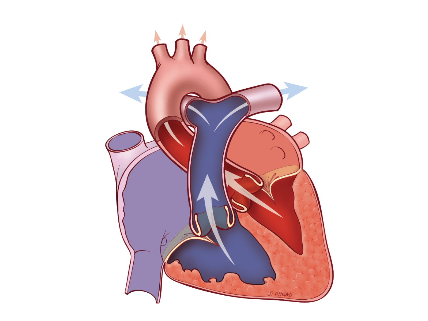
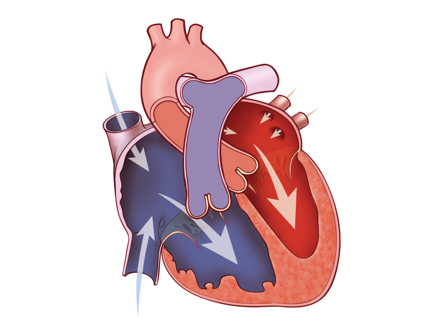
The heart is a muscular organ located in the center of the chest between the lungs. Your heart is about the size of your two hands when they are clasped together. The heart is a sophisticated pump, providing blood flow to all of the cells, tissues and organs in your body.
Normal, healthy heart valves
Human heart valves are remarkable structures. These tissue-paper thin flaps, or leaflets, attached to the heart wall undergo a constant “beating” as they open and close with each heart beat, day after day, and year after year. With each beat, the valves display their remarkable strength and flexibility.
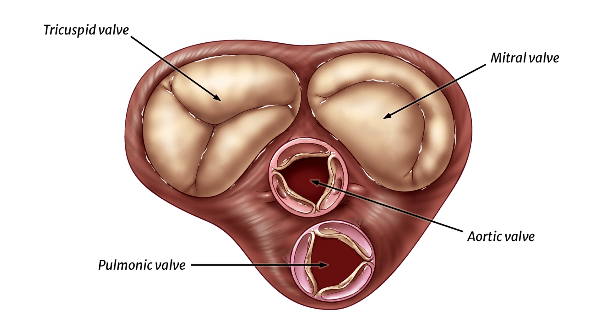
There are four cardiac valves. Two of the valves are referred to as the atrioventricular (AV) valves. They control blood flow from the atria to the ventricles. The AV valve on the right side of the heart is called the tricuspid valve; it sits between the right atrium and the right ventricle. The name “tricuspid” refers to the three leaflets making up the valve. The AV valve on the left side of the heart is called the mitral valve; the mitral valve controls blood flow between the left atrium and left ventricle. It has two leaflets. The other two cardiac valves, i.e., the aortic and pulmonic valves, each have three leaflets. They are outflow valves, regulating the flow of blood as it leaves the ventricles and the heart. The aortic valve serves as the “door” between the heart and the rest of the body. Every drop of blood ejected by the left ventricle must pass through the aortic valve. The aortic valve is located between the left ventricle and the ascending aorta, or upper portion of the aorta. The pulmonic valve is located between the right ventricle and the pulmonary artery. The mitral and tricuspid valves are substantially larger than the aortic and pulmonic valves.
The image below shows the shape and location of the four heart valves. The characteristic heart sounds (“lub-dub”) are caused by the closing of the heart valves; the first by closure of the mitral and tricuspid valves and the second by closure of the aortic and pulmonic valves.


Function of heart valves

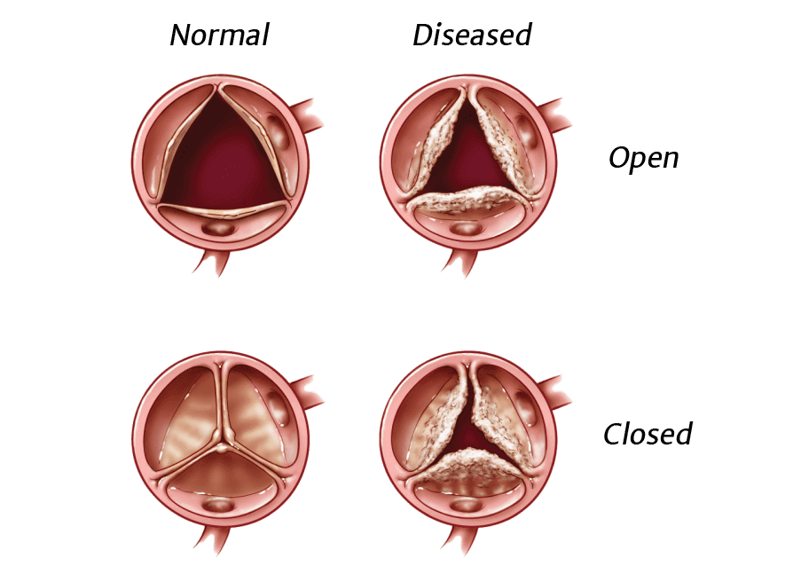
A normal, healthy valve would be one which minimizes obstruction and allows blood to flow freely in only one direction. It would close completely and quickly, not allowing much blood to flow back through the valve (backward flow of blood across a heart valve is called “regurgitation”). While a small amount of regurgitation, or leak, may be present and is well tolerated, severe regurgitation is always abnormal.
When a heart valve opens fully and evenly, blood flows through the valve in a smooth and even manner. When a valve does not open fully or evenly, blood flow through it becomes chaotic and turbulent. When a valve is narrowed or does not open fully, it is said to be “stenotic.”
Both regurgitation (a leak) and stenosis (a narrowing) increase the heart’s workload.
Defects and diagnosis

A variety of conditions can cause heart valve abnormalities, and there are many ways of determining if you have heart valve disease. Learn what happens with heart valve disease and how it is diagnosed.


Valve treatment

Diseased heart valves can be addressed in several ways. If repair or replacement becomes necessary, there are treatment options to consider. A team of medical specialists will be committed to your safety and comfort before, during and after your procedure.
How Edwards Lifesciences treats heart valve disease

Surgical valves
Surgical aortic valve replacement (SAVR) treatment options include replacing the valve through standard open-heart surgery or small-incision surgery. In both approaches, the surgeon removes the diseased valve and puts a new heart valve in its place.


